
From Particles to
New Research Possibilities
Explore published applications of magnetic and plasmonic materials.
Select a case to learn how it works and review the research findings.
Particle and Assembly Applications
Sensors, Tunable Optics, and Electrochemical Analysis of Structure
Magnetically Assembled Nanoparticles,
A Stronger Detection Signal
Siglec-15 Detection Using Ag@Fe₃O₄ Nanochains
Ag@Fe₃O₄ Nanochains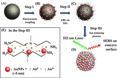
Rough Gold Surfaces
Enhance Raman Signals
SERS with Magnetic Particles Featuring a Concave Gold Shell
Fe₃O₄@Au Superparticles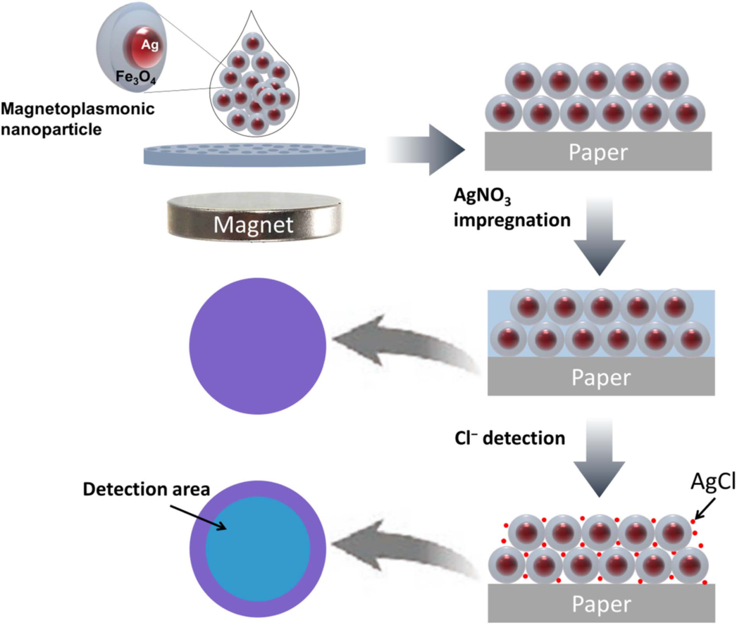
Chloride Ions in Water
Detected Through Color Change
Colorimetric Sensing with a Photonic Array on Paper
Ag@Fe₃O₄ Particle Array
Color Changes When Stretched
Flexible Sensor
Strain Sensing with a Magnetic-Plasmonic Particle Array
Ag@Fe₃O₄ Particles in an Elastomer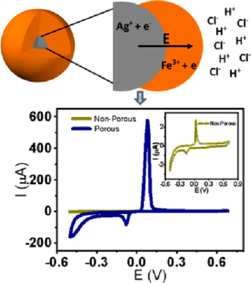
Paths into the Particle
Read Through Electrochemistry
Porosity and Molecular Access in Core-Shell Particles
Ag@Fe₃O₄ / Additional SiO₂ Coating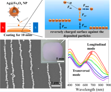
Tune Light with Polarization and
Interparticle Spacing
Polarization Dependence of Magnetically Assembled Nanochains
Ag@Fe₃O₄ NanochainsExtending to Nanowires and 1D Structures
Pre-commercial research platforms · Commercialization may be considered based on research demand
Case 07 is an assembled particle wire; cases 08–09 are directly synthesized Au@FeₓOᵧ nanowires.
One Immunoreaction,
Read by Raman and Fluorescence
Dual-Mode Detection of the CFP-10 Antigen
Assembled-particle MagPlas NW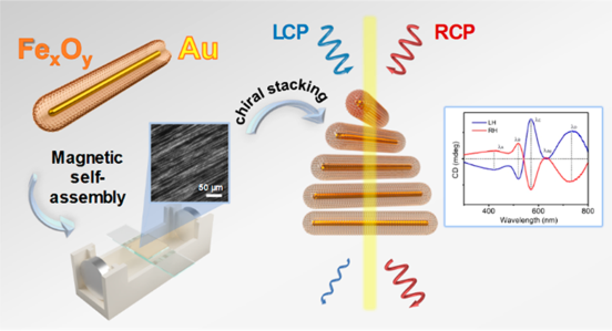
Twisted Nanowire Layers Form
A Chiral Optical Sensor
Biomimetic Bouligand Structures and Protein Response
Directly Synthesized Au@FeₓOᵧ NW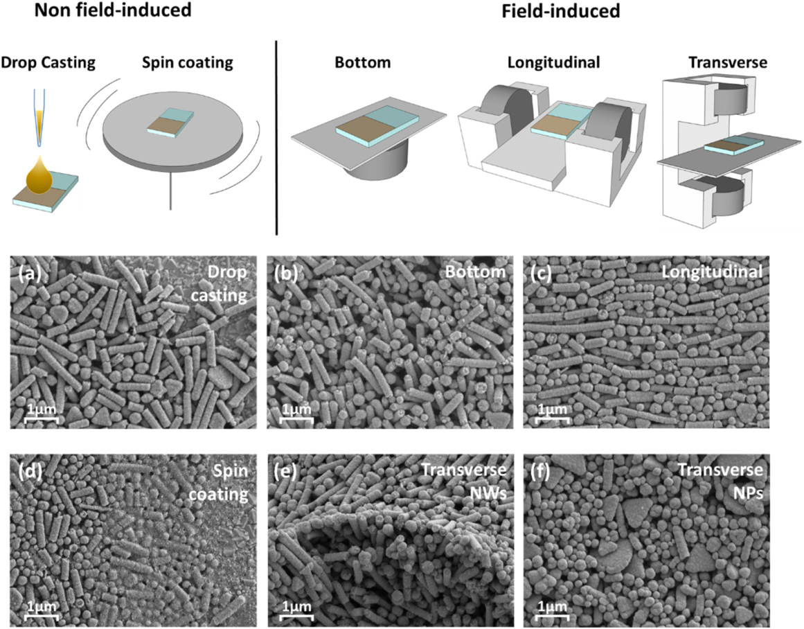
Nanowire Assembly
Improves Photoelectrode Performance
Water Oxidation with Ni Incorporation and Magnetic-Field Assembly
Ni-Incorporated Au@FeₓOᵧ NWMagnetically Assembled Nanoparticles,
A Stronger Detection Signal
Ag@Fe₃O₄ Nanochain-Based
Siglec-15 SPR Sensor
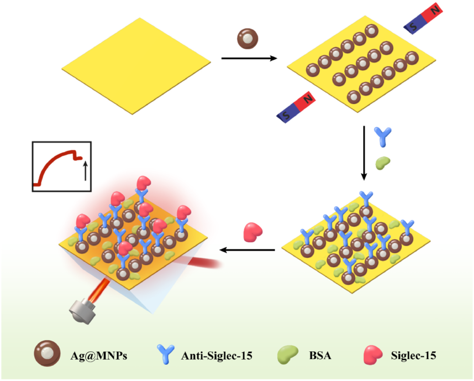
Magnetic-plasmonic nanoparticles were aligned in a row on a gold sensor surface. Adding this layer produced a stronger optical signal at the same protein concentration.
These values were obtained under the conditions reported in the paper. The Siglec-15 concentration response was evaluated over 1–500 ng/mL and should be distinguished from the calculated limit of detection.
How Do the Nanoparticles Contribute?
Magnetically Align on the Sensor Surface
The magnetic Fe₃O₄ shells are used to assemble Ag@MNPs into nanochains on a gold chip.
Capture the Target Protein with Antibodies
Polydopamine immobilizes the assembly and enables antibody attachment. The antibodies recognize Siglec-15, while BSA treatment reduces nonspecific binding.
Detect Binding Through an Optical Response
The sensor reads changes in the surface plasmon resonance (SPR) angle. The paper discusses optical coupling between the silver core and gold film as one possible source of signal enhancement.
How the Nanoparticle Layer Changes the Signal
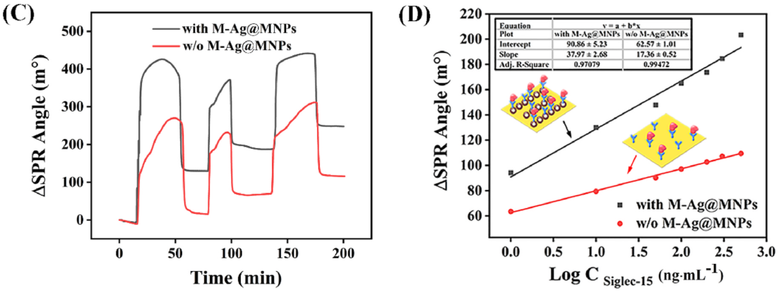
Stronger Response at the Same Concentration
The time-response plot on the left compares SPR signals during target detection. For 10 ng/mL Siglec-15, the paper reports an approximately 1.51-fold change in angle with nanochains.
Concentration Changes Also Produce an Optical Signal
On the right, the signal increases with the logarithm of Siglec-15 concentration. The sensor with nanochains showed a larger response over the concentration range tested.
Sensor Application of the Ag@Fe₃O₄ Platform
This study demonstrates particles that combine a silver core with a magnetic iron oxide shell. When designing a similar sensor with the SpinPlas Ag@MNP product line, surface condition, dispersion, magnetic-field assembly, and antibody immobilization conditions must be evaluated separately.
The identity of the particles in the paper and currently available products must be verified. The reported performance is from a complete research sensor and does not represent guaranteed performance of the particles alone or clinical validation.
Magnetic field-aligned Fe₃O₄-coated silver magnetoplasmonic nanochain with enhanced sensitivity for detection of Siglec-15
Read the Original Paper ↗Study Summary Based on the Abstract
This study presents a method for improving SPR sensor performance by using magnetically aligned Ag@MNP nanochains as the sensing layer on a gold chip. At a particle concentration of 80 ng/mL, refractive-index sensitivity, detection accuracy metrics, and the figure of merit improved compared with the gold chip alone. With Siglec-15 as a model target, a larger SPR angle change was observed at the same concentration, and the paper calculated a limit of detection of 1.36 pg/mL. This is an example of sensor design that combines magnetic assembly with plasmonic properties.
Rough Gold Surfaces
Enhance Raman Signals
SERS with Magnetic Particles Featuring a Concave Gold Shell
A rough, concave gold shell was formed on magnetic particles. The study created regions of concentrated light and used magnetic-field assembly to enhance molecular Raman signals.
Fe₃O₄@Au SuperparticlesThe results described here were obtained under the experimental conditions reported in the paper. They are distinct from guaranteed performance of products for sale.
How Does the Material Work?
Attach Gold Seeds
Coat magnetic iron oxide with PEI and attach small gold particles.
Grow a Rough Shell
Combine layer-by-layer assembly with gold-ion reduction to form a concave gold surface.
Assemble and Measure
Assemble the magnetic particles and compare the Raman responses of rhodamine B and graphene quantum dots.
Key Findings in the Paper
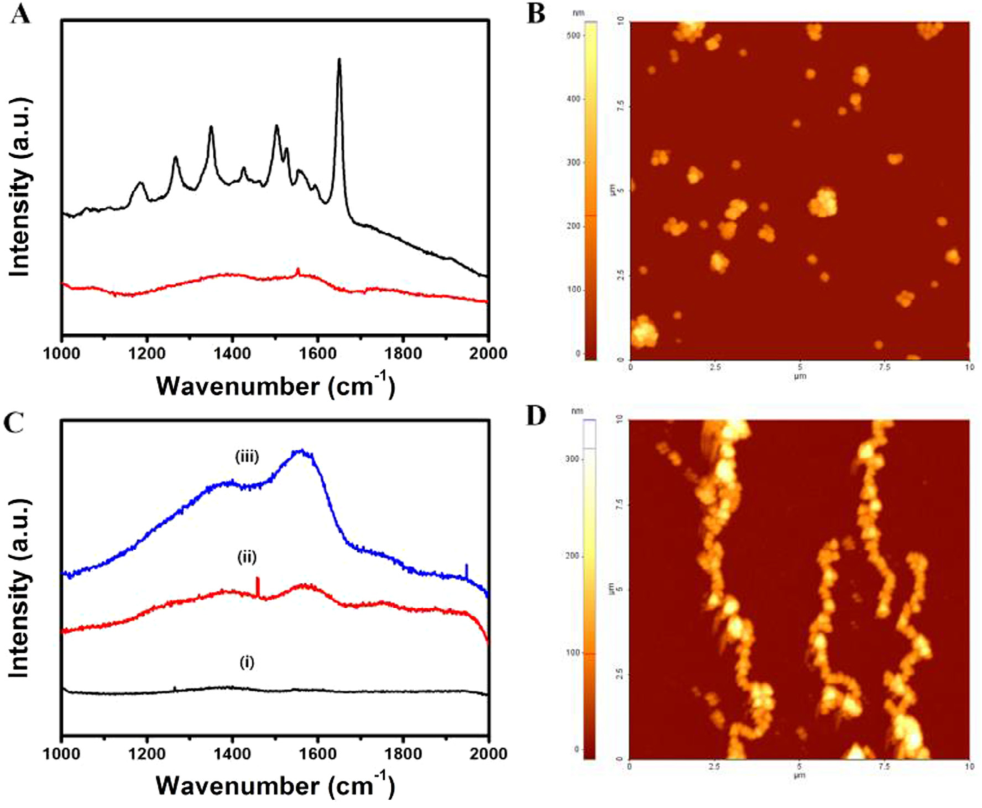
Figure 6 compares Raman signals with and without particles with a rough gold shell, as well as different assembly states. It highlights the difference between dispersed and assembled particles for graphene quantum dots.
Relevance to the SpinPlas Platform
The particles in this study have an Fe₃O₄ core and an Au shell. Their core-shell order and metal differ from current Ag-core, iron-oxide-shell products; this case is presented as an extension involving gold surfaces.
Preparation of concave magnetoplasmonic core-shell supraparticles of gold-coated iron oxide via ion-reducible layer-by-layer method for surface enhanced Raman scattering
Read the Original Paper ↗Study Summary Based on the Abstract
The study used an IR-LbL method to form a gold shell on magnetic iron oxide and control its thickness and roughness. The resulting superparticles were assembled with a magnetic field and their Raman signals measured, demonstrating the potential of SERS substrates that combine surface morphology and assembly.
Chloride Ions in Water
Detected Through Color Change
Colorimetric Sensing with a Photonic Array on Paper
When a nanoparticle array on paper encounters chloride ions, its color changes from purple to blue. This water-analysis study links the particles' structural color with AgCl precipitation.
Ag@Fe₃O₄ Particle ArrayThe sample preparation in the paper includes drying at 60 °C for 3 minutes. Figure 2 uses 250 mM AgNO₃, while the separately reported lowest LOD of 0.233 mM was obtained with 500 mM AgNO₃.
How Does the Material Work?
Form the Array
Create an amorphous photonic array of Ag@Fe₃O₄ particles on paper.
Add the Reagent
Impregnate the paper with AgNO₃ to provide Ag⁺ ions that react with chloride.
Read the Color Change
Observe the color and reflectance spectrum as AgCl forms.
Key Findings in the Paper
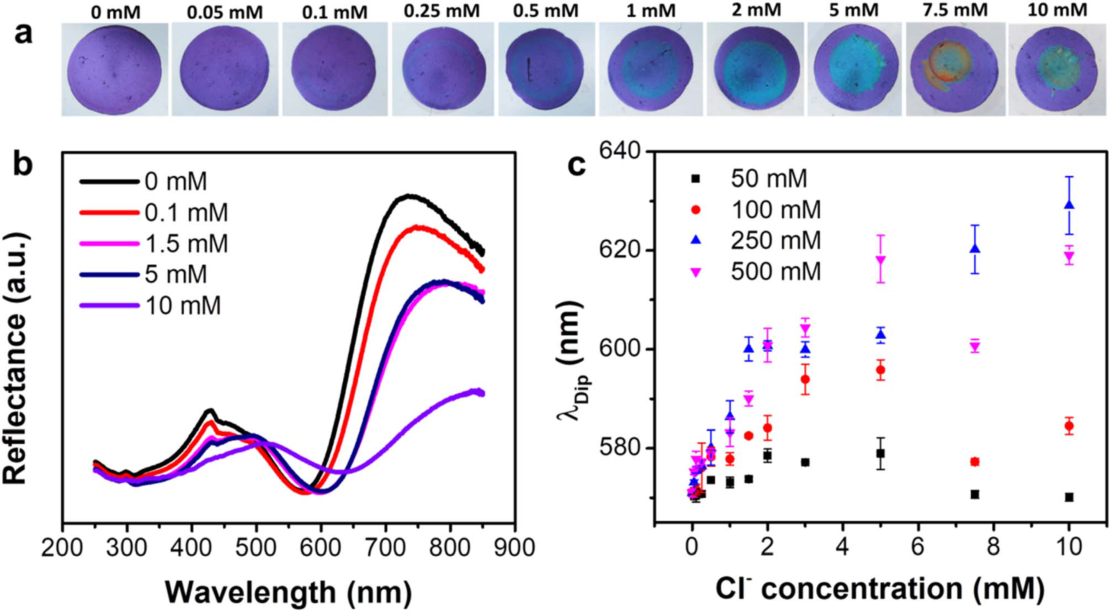
Figure 2 shows photographs and reflectance spectra at different chloride concentrations. Because the color response depends on particle size and AgNO₃ impregnation concentration, the particles alone do not guarantee the same response.
Relevance to the SpinPlas Platform
This particle-based application relates to the Ag@Fe₃O₄ product line. It additionally requires an array on a paper substrate and AgNO₃ impregnation; the nanoparticle dispersion itself is not a finished chloride test strip.
Magnetoplasmonic photonic arrays for rapid and selective colorimetric detection of chloride ions in water
Read the Original Paper ↗Study Summary Based on the Abstract
AgNO₃ was introduced into an amorphous photonic array of Ag@Fe₃O₄ particles, changing the local refractive index through a precipitation reaction with chloride ions. Chloride was detected through color and reflectance spectra, and interference from other ions and use with real samples were evaluated.
Color Changes When Stretched
Flexible Sensor
Strain Sensing with a Magnetic-Plasmonic Particle Array
When a nanoparticle array is combined with an elastomer, its color changes as it is stretched or bent. This materials study represents strain as a visible optical signal instead of an electrical one.
Ag@Fe₃O₄ Particles in an ElastomerThe results described here were obtained under the experimental conditions reported in the paper. They are distinct from guaranteed performance of products for sale.
How Does the Material Work?
Assemble the Particles
Use a magnetic field to form an amorphous photonic array.
Combine with an Elastomer
Combine the array with a flexible medium to make a deformable film.
Visualize Movement
Read changes in interparticle interactions and structural color as the material deforms.
Key Findings in the Paper
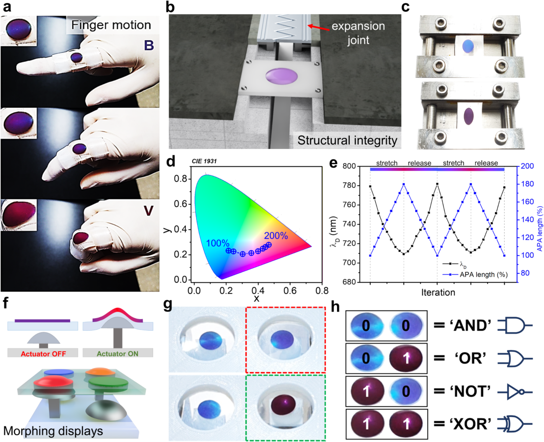
Figure 6 shows examples of finger-joint movement, material deformation, and display devices. It also presents optical responses over repeated stretching and relaxation, demonstrating how deformation appears as a color change.
Relevance to the SpinPlas Platform
This is an application of assembled spherical Ag@Fe₃O₄ particles, not a nanorod-based study. Product application requires optimization of particle size, assembly, and elastomer fabrication conditions.
Mechanochromic strain sensor by magnetoplasmonic amorphous photonic arrays
Read the Original Paper ↗Study Summary Based on the Abstract
An amorphous photonic array was formed by magnetically assembling Ag@Fe₃O₄ core-shell particles and combining it with an elastomer. The color-change range was tuned by particle size; a distinct blue-to-red transition was observed with 176 nm particles. The study demonstrated applications for visualizing joint movement and structural deformation.
Paths into the Particle
Read Through Electrochemistry
Porosity and Molecular Access in Core-Shell Particles
Core-shell particles that look similar from the outside can differ in how readily substances reach their interiors. This study compares shell accessibility using the redox response of a silver core.
Ag@Fe₃O₄ / Additional SiO₂ CoatingThe results described here were obtained under the experimental conditions reported in the paper. They are distinct from guaranteed performance of products for sale.
How Does the Material Work?
Prepare Comparison Particles
Compare Ag@Fe₃O₄ particles with particles that have an additional SiO₂ coating.
Apply Potential
Vary the potential and observe the electrochemical responses of the silver core and iron oxide shell.
Compare Structure and Response
Relate current changes to electron-microscopy observations to interpret shell accessibility.
Key Findings in the Paper
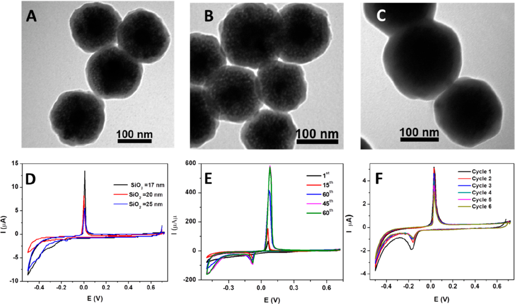
Figure 4 shows the relationship between SiO₂ shell thickness and electrochemical response. The paper reports an Ag oxidation peak current of 400:3.2 μA for the porous shell versus the SiO₂-coated shell. This is not a ratio of porosity.
Relevance to the SpinPlas Platform
This study helps explain shell accessibility in Ag@Fe₃O₄ products and the effect of additional coatings. It may inform catalyst or mass-transfer platform design, but the paper does not validate drug-delivery efficacy.
Electrochemical Investigation of Porosity in Core–Shell Magnetoplasmonic Nanoparticles
Read the Original Paper ↗Study Summary Based on the Abstract
Ag@Fe₃O₄ and Ag@Fe₃O₄@SiO₂ models were used to distinguish redox reactions at the core and shell. Differences in molecular access through the shell were identified from current responses and compared with electron-microscopy results. This is an example of using electrochemistry to analyze multicomponent nanoparticle structures.
Tune Light with Polarization and
Interparticle Spacing
Polarization Dependence of Magnetically Assembled Nanochains
The same nanoparticles can respond to light differently when assembled in a row. This optical-film study tunes resonance wavelengths through polarization direction and interparticle spacing.
Ag@Fe₃O₄ NanochainsThe results described here were obtained under the experimental conditions reported in the paper. They are distinct from guaranteed performance of products for sale.
How Does the Material Work?
Set the Spacing
Adjust the iron oxide shell thickness to change the spacing between silver cores.
Assemble Nanochains
Use a magnetic field to form a one-dimensional particle array on a glass substrate.
Adjust Polarization
Change the polarization direction of incident light and compare the array's resonance wavelengths.
Key Findings in the Paper
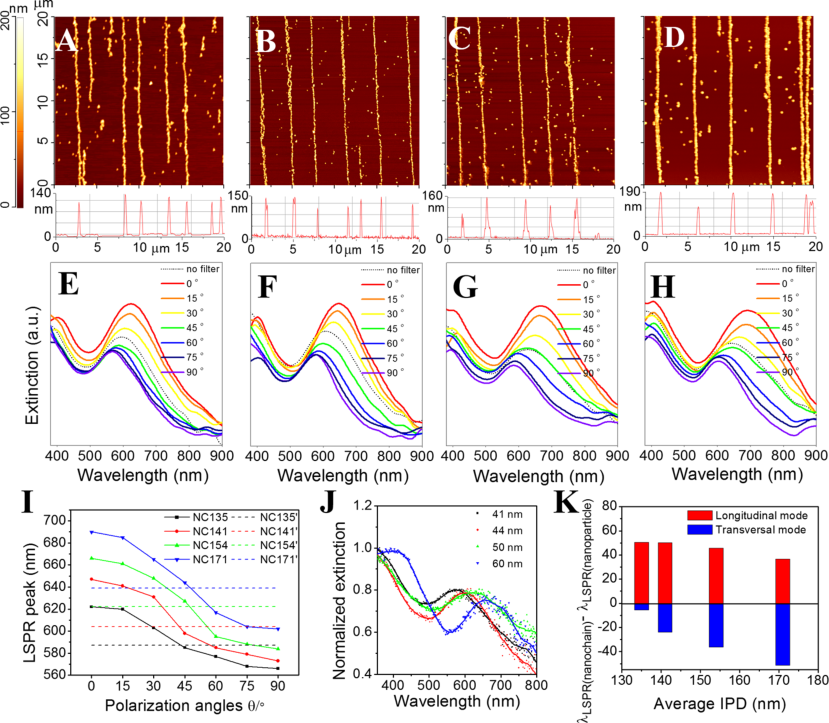
Figure 3 shows array morphology at different shell thicknesses and polarization-dependent extinction spectra. The paper interprets far-field optical coupling as important because the spacing between silver cores is relatively large.
Relevance to the SpinPlas Platform
These nanochains are assembled from spherical Ag@Fe₃O₄ particles. They are distinct from directly synthesized nanorods or nanowires; shell thickness and assembly conditions must be tailored to the desired optical response.
Tuning Plasmon Resonance in Magnetoplasmonic Nanochains by Controlling Polarization and Interparticle Distance for Simple Preparation of Optical Filters
Read the Original Paper ↗Study Summary Based on the Abstract
Ag@Fe₃O₄ particles were assembled into large-area nanochains, and plasmon resonance was analyzed as a function of polarization angle and interparticle spacing. The shell provides magnetic functionality and sets the spacing between cores. This work informs the design of tunable optical filters and anisotropic optical platforms.
One Immunoreaction,
Read by Raman and Fluorescence
Dual-Mode Detection of the CFP-10 Antigen
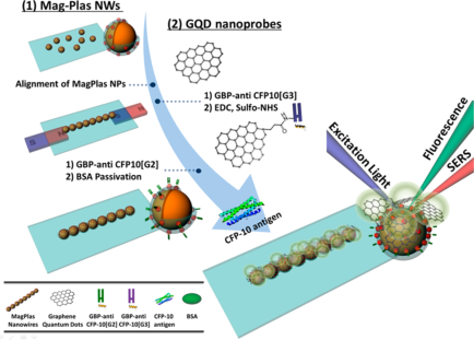
Antibodies and graphene quantum dots were attached to particles assembled in a line by a magnetic field. This study visualized target binding by fluorescence and quantified it through Raman signals.
Assembled-particle MagPlas NW · Pre-commercial research platformThe results described here were obtained under the experimental conditions reported in the paper. They are distinct from guaranteed performance of products for sale.
How Does the Material Work?
Assemble in One Dimension
Magnetic particles coated with gold are assembled by a magnetic field. The paper calls this assembled structure MagPlas NW.
Form a Sandwich Assay
Different antibodies are immobilized on the array and graphene quantum dots to capture the target antigen between them.
Read Two Signals
Fluorescence visually confirms binding, while SERS is used to analyze antigen concentration.
Key Findings in the Paper
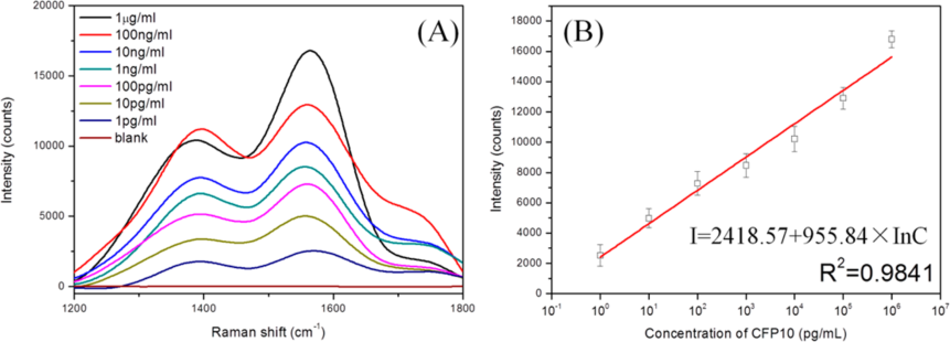
Figure 6 shows SERS spectra and calibration relationships at different CFP-10 concentrations. The concentration range compared is 1 pg/mL–1 μg/mL; the calculated limit of detection reported in the paper should be distinguished from this measurement range.
Pre-commercial Research Platform
This is a pre-commercial research platform. It is a wire-like structure assembled from Fe₃O₄@PEI–Au particles, differing in fabrication method and core-shell composition from the directly synthesized Au@FeₓOᵧ nanowires in cases 08 and 09.
Dual-Mode SERS-Fluorescence Immunoassay Using Graphene Quantum Dot Labeling on One-Dimensional Aligned Magnetoplasmonic Nanoparticles
Read the Original Paper ↗Study Summary Based on the Abstract
Graphene quantum dots served as dual Raman and fluorescence labels, and a sandwich immunoassay was assembled on one-dimensional magnetic-plasmonic particles. Using CFP-10 as a model antigen, the study observed fluorescence imaging and concentration-dependent SERS responses.
Twisted Nanowire Layers Form
A Chiral Optical Sensor
Biomimetic Bouligand Structures and Protein Response
Nanowires with a gold core and magnetic iron oxide shell were stacked at different orientations. The non-chiral material formed a helical structure with distinct responses to circularly polarized light, and changes were observed after adding a protein.
Directly synthesized Au@FeₓOᵧ NW · Pre-commercial research platformThe results described here were obtained under the experimental conditions reported in the paper. They are distinct from guaranteed performance of products for sale.
How Does the Material Work?
Synthesize Nanowires
Produce Au@FeₓOᵧ nanowires using a one-step solvothermal process.
Rotate and Stack
Rotate and stack magnetically aligned layers at set angles to form a helical structure.
Measure the Chiral Response
Measure circular dichroism (CD) and compare peak shifts after introducing BSA.
Key Findings in the Paper
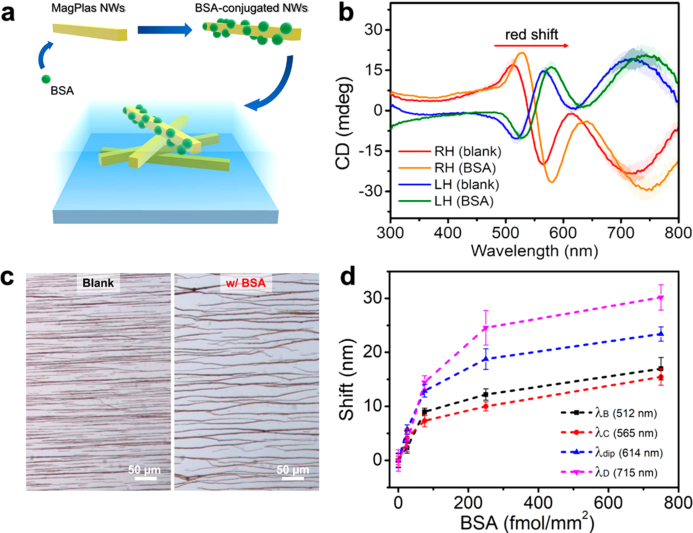
Figure 6 presents CD spectra and concentration-dependent peak shifts after BSA was introduced to the top layer. This is a study of structure-based chiral optical responses; it does not demonstrate clinical diagnostic performance or universal protein selectivity.
Pre-commercial Research Platform
This is a pre-commercial nanorod and nanowire research platform. Commercialization may be considered based on research demand. Its core metal and shape differ from the spherical Ag@Fe₃O₄ products currently for sale.
One-Pot Synthesis of Magnetoplasmonic Au@FexOy Nanowires: Bioinspired Bouligand Chiral Stack
Read the Original Paper ↗Study Summary Based on the Abstract
One-step-synthesized Au@FeₓOᵧ nanowires were magnetically aligned to create structural color. The study then used rotational stacking to make a Bouligand-type chiral film and observed circular dichroism and optical responses to the model protein BSA.
Nanowire Assembly
Improves Photoelectrode Performance
Water Oxidation with Ni Incorporation and Magnetic-Field Assembly
Performance depends on both catalyst composition and its arrangement relative to light. This study improved light absorption and water oxidation by incorporating Ni into magnetic nanowires and adjusting their orientation with a magnetic field.
Ni-incorporated Au@FeₓOᵧ NW · Pre-commercial research platformThe representative values are from the paper's abstract and conclusion. They refer to photoelectrode current density, not hydrogen production or guaranteed performance of a particle powder.
How Does the Material Work?
Adjust Composition
Incorporate Ni into Au@FeₓOᵧ nanowires to modify the surface properties.
Magnetic-Field Assembly
Adjust the nanowire orientation on the electrode by changing the magnetic-field direction.
Evaluate the Photoelectrode
Compare electrode performance by measuring photocurrent, light-conversion efficiency, and impedance.
Key Findings in the Paper
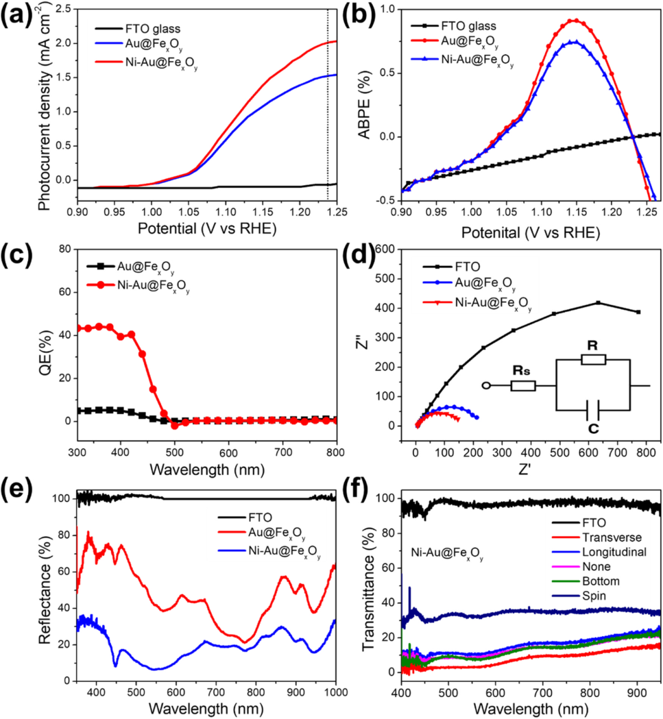
Figure 7 compares photoelectrochemical and optical responses of Au@FeₓOᵧ and Ni-incorporated nanowire electrodes. The paper reports that the representative photocurrent density increased from 1.52 to 2.01 mA/cm² at 1.23 V vs. RHE.
Pre-commercial Research Platform
This is a pre-commercial nanorod and nanowire catalyst research platform. Any future commercialization assessment should consider Ni incorporation conditions, fabrication of assembled electrodes, and photoelectrochemical evaluation.
Tuning assembly of Ni-doped Au@FexOy magnetoplasmonic nanowires for optimizing photochemical water splitting
Read the Original Paper ↗Study Summary Based on the Abstract
Ni-incorporated Au@FeₓOᵧ nanowires were synthesized, and their assembly on an electrode was controlled with a magnetic field. Controlling light absorption and surface structure improved photoelectrochemical performance for water oxidation, showing that electrode orientation is an important design variable alongside material composition.
Mechanochromic Sensing Patch
Flexible Sensor Patch That Reads Substrate Strain Through Color
How it works
- Form a magnetic-plasmonic particle array based on SP-MPB-COOH.
- Implement the particle array on a flexible elastomer substrate.
- Observe the optical response as a color change when the substrate is stretched or bent.
Potential applications
- Visualize Local Strain in Structures
- Monitor Repeated Deformation in Flexible Components
- Visualize Joint Movement and Motion
- Camera-Based, Non-contact Readout
Standard product specifications have not yet been finalized. Customer joint evaluation is underway to assess substrate surfaces, strain ranges, cycling conditions, and readout methods.
Inquire About a Joint Evaluation →No standard product specifications are currently available. Color-change magnitude, sensing range, cycle life, and environmental durability will be assessed through joint evaluation for each application. Results reported in papers are not guaranteed specifications for this prototype.